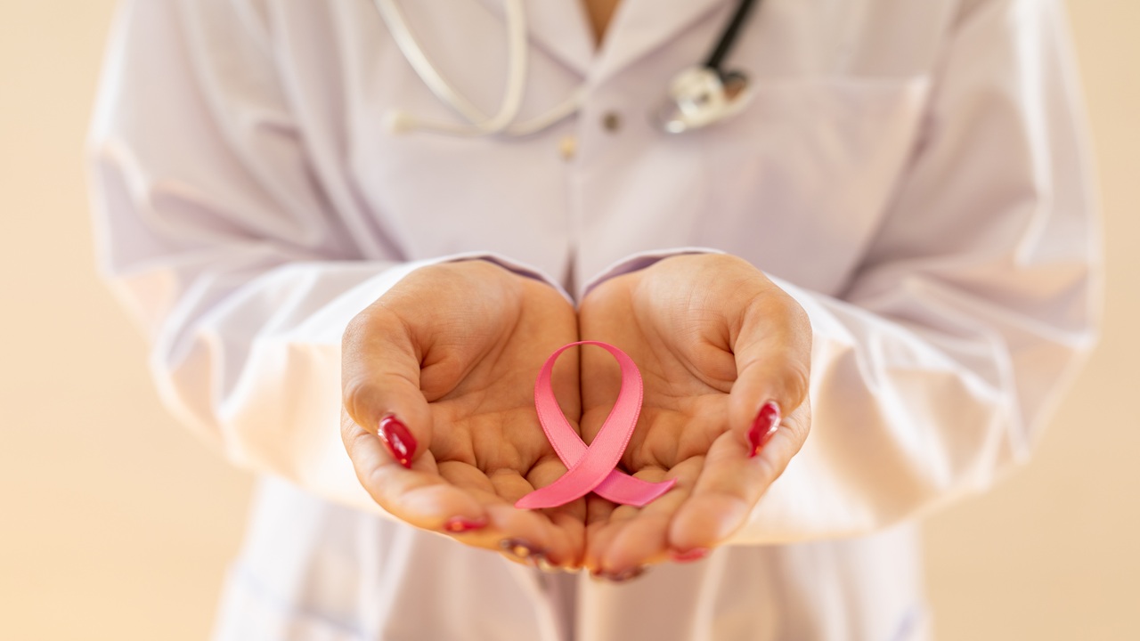
Discover the challenges in screening and after a biopsy when you have dense breasts

Dense Breast: Risk, Screening Challenges and What Happens After a Biopsy
October 15, 2025
Beyond the issue of detection, dense breast tissue slightly increases the risk of developing breast cancer over a woman’s lifetime. The higher the density, the higher the risk tends to be. Women with extremely dense breasts (category D, extremely dense, may make tumors difficult to see) have about a 4–6 times higher risk of breast cancer compared to women with almost entirely fatty breasts (category A).
In more practical terms, this roughly translates to about a 2‑fold higher risk compared to an average-density woman.
The reasons aren’t fully understood, but one factor may be that dense breasts have more glandular cells which can undergo cancerous changes. However, it’s very important to keep this risk in perspective: most women with dense breasts will never develop breast cancer. Breast density is just one of many risk factors – other factors like age, family history, and certain genetic mutations can contribute to risk as much or more than density. So having dense breasts doesn’t mean you’re destined to get cancer; it simply means we need to be a bit more vigilant with screening.
Challenges in Screening and After a Biopsy
Because dense breasts can hide cancer, there are a few practical challenges that women with dense tissue should be aware of:
- Mammogram limitations: A standard 2D mammogram might not catch every cancer in dense tissue. Radiologists know to be cautious – something that looks “normal” on a dense breast image could be hiding a small tumor. This is why, in many cases, radiologists may recommend additional imaging (like an ultrasound or MRI) if you have dense breasts, even when your mammogram doesn’t show a clear abnormality. Mammography is still essential, but in some women it may not be sufficient on its own. An MRI is normally the best breast exam for dense breasts.
- False alarms and biopsies: Dense tissue not only hides cancers, it can also make normal tissue look suspicious. Women with dense breasts are more likely to get called back after a mammogram for further tests because something on the initial image is hard to interpret. Many times these callbacks do not end up being cancer – they could be overlapping dense tissue or benign findings. As a result, dense breasts slightly increase the chance of undergoing a biopsy that turns out benign. This can be stressful, but it’s done out of caution. A benign result provides peace of mind; a false positive doesn’t increase the risk of dying of cancer, but it does mean extra tests.
- Follow‑up after a biopsy:
- If a biopsy is benign, short‑term follow‑up imaging (e.g., at 6 months) is often recommended to ensure stability.
- If a biopsy finds cancer, dense breasts can influence next steps. Your team might order a breast MRI to better define the extent of disease, since MRI can see through dense tissue more effectively.
- During any biopsy, the radiologist places a tiny clip/marker at the biopsy site so future imaging shows exactly where it was. In very dense tissue, sometimes MRI‑visible markers are used to ensure accurate long‑term tracking.
FAQ: Dense Breasts, Risk, and Screening Challenges
Does having dense breasts mean I will get breast cancer?
No. Most women with dense breasts do not develop cancer. While density slightly increases risk, other factors—like age, genetics, and family history—are just as important.
Why are mammograms less accurate with dense breasts?
Because dense tissue and tumors both appear white on a mammogram. This can hide small cancers, a problem known as the masking effect. MRIs are the best breast exam for dense breast.
Can dense breasts lead to more false alarms?
Yes. Dense tissue can look suspicious on imaging, even when it’s normal. This leads to more callbacks and benign biopsies, especially in categories C and D.
What happens after a benign breast biopsy?
You’ll usually have a short-term follow-up (often a mammogram or ultrasound in 6 months) to confirm that the area is stable.
Do dense breasts change biopsy procedures?
The procedure is the same, but radiologists may use a special clip or MRI-visible marker to help track the biopsy site over time, especially in extremely dense tissue.
Join the Revolution in Women’s Health
As VM-1 sets a new standard for post-biopsy care, we’re not just improving technology—we’re improving lives.
About VizMark
Our team, led by radiologist and women’s health advocate Dr. Michael T. Nelson, created VM-1 improve imaging under MRI necessary for high risk patients. Through years of research and collaboration, we’ve designed a marker that eliminates the challenges of traditional solutions, offering better outcomes for patients and providers alike.

Dr. Michael Nelson
Founder and primary inventor, Dr. Nelson is a Board-Certified Radiologist, former U.S. Navy Flight Surgeon, and Professor of Radiology at the University of Minnesota. A leader in Mammography, Interventional Radiology, and Women’s Health, he co-founded the Jane Brattain Breast Center at Park Nicollet Medical Center.
Dr. Michael Nelson
Founder, Chief Medical Officer, and radiology expert

Kim Nelson
Accomplished senior executive and board member with 30+ years in high-tech, leading business growth, revenue expansion, and successful exits, including roles at Oracle and Primus Knowledge (IPO).
Kim Nelson
CEO with a proven track record of driving innovation

Tom Murphy
With 30+ years in medical device sales, operations, and management, Mr. Murphy has deep expertise in the tumor marker space, leading operations and sales at Mermaid Medical, Argon Medical, and Angiotech.
Tom Murphy
VP of Operations, tumor marker specialist

Steve Karel
A biotech senior executive with startup experience, Mr. Karel has held roles as CEO, CFO, and SVP of Business Development. He brings expertise in finance, fundraising, corporate development, and R&D, strengthening VizMark’s operations and growth.
Steve Karel
CFO with deep expertise in medical finance
Suite 150, 10580 Wayzata Blvd, Plymouth, MN 55305